
Understanding Your Digital Eye Images and Results

Have you ever sat in the exam chair and seen a detailed, colorful picture of your own eye? Your doctor might point things out, but later, you think, “What was I really looking at?”
But these images are not confusing on purpose. Let us talk about what these pictures are and how to understand your results. It is simpler than you might think.
A Snapshot of Your Inner Eye
First, what are we seeing? This is not a simple photo. It is a high-tech image of the inside wall at the back of your eye, called the retina.
Your retina is like the film in a camera. It captures light and sends the picture to your brain. When your doctor takes a digital image, they get a clear view of this delicate area. They can see your optic nerve, blood vessels, and the macula (your central vision spot). This technology, known as digital retinal imaging, lets them see details they could not spot with a simple glance.
Why Your Doctor Takes These Pictures
You might wonder, “Is this really needed?” For modern eye care, the answer is yes. Here is why.
These images give your doctor a permanent record. It is like a baseline map of your eye’s health. At your next visit, they can compare the new images to the old ones. This helps them spot the tiniest changes over time. Catching a small change early is everything. It is the best way to stay ahead of potential issues.
They can reveal early signs of conditions long before you notice any symptoms. This includes things like glaucoma, diabetic retinopathy, or macular degeneration. It is a key part of preventative care.
Making Sense of What You See
So, you are looking at an image with swirls of orange and red. What does it all mean?
Instead of diagnosing yourself, think in terms of patterns. Your doctor is looking for clear, consistent textures and colors.
Healthy signs include a uniform background color and blood vessels that look like clear, branching lines.
Areas for watchfulness might look unusually dark, pale, or blurred. This could signal thinning, fluid, or other changes.
Your job is not to interpret these signs. Your job is to understand why the image is so valuable. It is a tool for early detection.
Your Results: A Chance for Conversation
When you get your results, use them as a conversation starter. Ask your doctor simple questions:
“Can you show me what a healthy part looks like on my image?”
“Did you see anything you want to watch?”
“What does this mean for my long-term eye health?”
This turns a technical report into a personal understanding. You become a partner in your own health.
The Goal Is Your Long-Term Vision
These digital images might feel like a lot of technology. But see them for what they are: a gentle, non-invasive form of protection. They give you and your doctor the clearest possible picture of your eye health year after year.
At your next exam, look at that image with curiosity. Ask your doctor to explain your personal map. It is one of the best steps you can take to protect your sight for all the moments that matter.
To understand your digital eye images and results, visit Custom Eyes Optical at our Selden, New York, office. Call (631) 230-6230 to book an appointment today.
https://www.sciencedirect.com/topics/engineering/iris-image
https://resources.altium.com/p/what-eye-diagram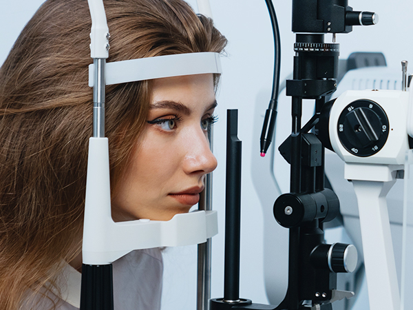

MAP
Powered by: 